Fallen Arches

How Fallen Arches Can Cause Low Back Pain

The human body is an intricate machine; everything is connected in one way or another. This means that when one part is not functioning as it should it can throw everything out of balance. The relationship between the feet, back, and hips is significant, so when there is a problem with feet it can cause pain in those areas. Fallen arches is a foot problem that is a very common cause of lower back pain. However, many people who have fallen arches don’t even realize that they have an issue.

What are Fallen Arches?

There are more than 100 ligaments and muscles in the foot that stabilize the bones and keep everything in the proper position. Ligaments prevent joints from extending beyond their intended limits. They contain elastin which allows them to act much like a rubber band, stretching with movement or flexion then returning to their normal length when the joint returns to its resting position.

There are variety of things that can happen that cause those ligaments to stretch, causing them to lose their ability to support the foot as they should. This can lead to pes planus (flatfeet), also known as fallen arches.

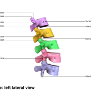
Most people don’t realize that there are actually three arches in the foot, not just one: lateral, medial, and anterior. They act as shock absorbers for the body. When they don’t function as they should, don’t absorb the shock of movement and activity, it puts added wear and tear on the body. While foot, knee, hip, and back pain is common with fallen arches, it also is a major cause of impaired functioning of the lower extremities. It can also lead to balance problems.

Causes of Fallen Arches

The primary cause of fallen arches is improper footwear. Shoes that do not adequately support your foot and arch can lead to structural problems.

Other causes of fallen arches include:

  • Tendons in the foot that are torn or stretched
  • Certain health conditions including
    • Rheumatoid arthritis
    • Pregnancy
    • Diabetes
    • Obesity
    • Aging
  • Structural abnormalities that are present from birth
  • Inflammation, deformity, or damage of the posterior tibial tendon that runs from the middle of the arch to the lower leg, running along the ankle
  • Nerve issues
  • Bones that are dislocated or broken

Recognizing the symptoms of fallen arches and knowing that you have a problem can help to prevent the pain but can also arrest further damage to the foot.

Do you have Fallen Arches?

While many people with fallen arches experience no symptoms or problems, others do. If you experience any of these symptoms of flat feet, it should prompt a visit to your doctor.

  • Pain in the foot, knee, leg, hip, or lower back
  • Feet that easily get tired
  • Difficulty with balance
  • Swelling on the bottom of the foot
  • Pain or achiness in the foot, particularly in the heel or arch
  • Certain movements. such as standing on tip toe, are difficult

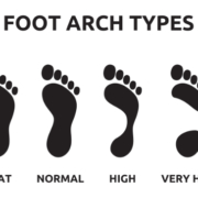
There is a quick, simple test that you can do at home to see if you have fallen arches:

  1. Wet your feet
  2. Stand on a flat, hard surface, such as a floor or concrete pad (you need to be able to see your footprint) or walk in soft soil or sand
  3. Look at your footprints. If you see a footprint that shows the heel and ball of your foot with a thin, curved imprint running along the outside, then your foot structure is normal. If you can see the imprint of your entire foot then you probably have fallen arches.

The problem with fallen arches is that the damage cannot really be reversed once it begins. However, the progression can be stopped with proper treatment.

What are the Treatments for Fallen Arches?

The treatment for fallen arches depends on the cause and severity of the condition. If it isn’t causing pain or other problems, the doctor may elect to do nothing.

However, if there is pain or mobility issues present, treatments may include bracing, prescribing NSAIDs for pain and inflammation, and even surgery in some cases of torn tendons or broken bones. An increasingly popular treatment for fallen arches is chiropractic care. This natural, non-invasive therapy treats the whole body so that it addresses the cause of the condition as well as associated risk factors and helps patients manage the pain, greatly improving their quality of life.

Visit www.ocwc.ca for more health and wellness tips!

Lumbar Spine Anatomy

Anatomy 101 – The Lumbar Spine: What Chiropractic Patients Need to Know

The lumbar spine, or lower back, take a lot of stress and strain throughout the day. The spine provides stabilization for the entire body and the lumbar spine bears the brunt of much of the movement and activity. This wear and tear shows up as lower back pain in many people. The American Chiropractic Association estimates that 31 million Americans have low back pain at some point in their lives. In fact, the leading cause of disability is low back pain – not only in the U.S. but worldwide.

What is the Lumbar Spine?

Located in the lower back area, the lumbar region is the portion of the spine that begins below the shoulder blades, about five or six inches, at the lordosis (where it curves in toward the abdomen). There are five vertebrae that make up the lumbar spine, L1 through L5 – which also happen to be the largest unfused vertebrae in the entire spinal column. The lower vertebrae bear more weight than the upper vertebrae so they are constructed to be sturdier, but are also more prone to injury and degradation.

The L5 vertebra meets the S1 (the sacrum) at the lumbosacral joint. It is this joint that provides most of the range of motion including rotational flexibility of the hips and pelvis, allowing them to swing during movement such as running and walking.

The spinal cord ends at around the L1 and many nerve roots that originate at the spinal cord form what is called the cauda equine. These nerves travel down the spine, branching out to the legs, buttocks, and feet. The appearance resembles a horse’s tail, hence the name. Because the spinal cord does not extend into the lumbar area, the chance of a problem with the lower back resulting in paralysis or damage to the spinal cord is very rare.

Lower Back Pain

The most common causes for lower back pain are soft tissue injuries and mechanical problems. Either of these can damage the intervertebral discs, compress the nerve roots, and inhibit correct function of the spinal joints so that they don’t move properly. Lower back pain is most commonly caused by a pulled or torn muscle or ligament, known as a sprain or strain. These injuries can have a number of causes, including:

  • Prolonged improper posture
  • Improper lifting of something heavy (without using your legs)
  • Sports injuries, particularly those where the player sustains high impact or where a lot of twisting is involved
  • Lifting while twisting the spine
  • A fall or movement that occurs suddenly and puts a great deal of stress on the lumbar area

Chiropractic Care for the Lumbar Spine

Chiropractic care is a very popular, effective treatment for lower back pain. A chiropractor can help patients manage their back pain through spinal adjustments and lifestyle advice. They may recommend supplements, exercise, and dietary changes. The whole-body approach to chiropractic makes it an optimal choice for many people, especially those who don’t want to take medications or undergo invasive procedures.

The chiropractic treatment itself usually involves spinal manipulation and mobilization. Spinal manipulation is the technique that most people think of when they think of chiropractic. It involves a short lever, high velocity arm thrust applied directly to the vertebra that is injured or is causing the pain. This technique typically relieves pain, improves the function of the spine, and restores the back’s range of motion. Mobilization is a low velocity movement performed by the chiropractor to manipulate the area, stretching and moving the joints and muscles.

These techniques typically restore range of motion and flexibility to the spine while reducing pain. For people with chronic lower back pain, chiropractic care can be a game changer.

Visit www.ocwc.ca for more health and wellness information! Call (780)455-2112 to book with one of our chiropractic doctors.

Cervical Spine Anatomy

Anatomy 101 – The Cervical Spine: What Chiropractic Patients Need to Know

Neck pain is one of the most common sources of pain and chronic pain worldwide. According to the International Association for the Study of Pain, each year, around 30% to 50% of the general population experiences neck pain and around 15% will, at some point in their lives, have chronic neck pain. Women seem to experience it more often than men and it is most prevalent at around middle age. Neck pain can be debilitating, impacting a person’s home life as well as their work performance. It can also trigger migraines and limit range of motion. Understanding the cervical spine is integral in understanding how to manage pain in that area.

What is the Cervical Spine?

There are seven vertebrae that make up the cervical spine: C1 through C7. They protect the spinal cord and are part of the system that makes up the neck.

C1 is located at the base of the skull and C7 sits at the beginning of the thoracic spine. While C1 is the smallest vertebrae, each subsequent one is slightly larger as you move down the spine. This is necessary because the farther down the spine, the more weight it must bear.

The vertebrae C3 through C6 are called “typical vertebrae.” Like other vertebrae in the spine, they have a similar construction. The top vertebrae, C1 and C2 are “atypical vertebrae.” Their construction is somewhat different from typical vertebrae due to their specialized function and location.

The atlas, C1, is the only vertebrae that has more of a ring shape than a shape resembling a vertebra. It is what connects the skull to the spine and is responsible for about half of the head’s backward and forward range of motion.

The axis, C2, is the second vertebra and has a special construction that connects it to C1 at the atlanto-axial joint. It is responsible for around half of the head’s rotation. The vertebra prominens, C7, is much larger than the vertebrae that sit above it and its shape is different to facilitate its connection to T1, at the beginning of the thoracic spine.

Neck Pain

The cervical spine has several critical functions. It houses the spinal cord and protects it, supports the head and facilitates its movement, and facilitates the flow of blood to the brain.

The human head is around 10 to 13 pounds and the cervical spine, along with an intricate network of muscles, tendons, and ligaments support it. This is what also allow flexibility to the head so that it can move up and down, backwards and forwards, rotational, and side bending. This job alone puts a great deal of stress on the neck and can lead to neck pain. Common causes of neck pain include:

  • Whiplash (whipping the head forwards and then backwards very suddenly)
  • Degenerative disc disease
  • Pinched nerve
  • Age related conditions
  • Spinal stenosis
  • Sleeping in certain positions
  • Neck strain
  • Osteoarthritis
  • Keeping the neck in one position too long, such as looking down at a mobile device
  • Herniated disc
  • Neck injury
  • Fibromyalgia

Chiropractic Care for the Cervical Spine

A chiropractor will typically treat a patient with neck pain using cervical spinal manipulation, cervical spinal mobilization, or a combination of the two techniques. Cervical spinal manipulation is what most people think of regarding chiropractic treatment. It involves brief, quick thrusts that focus on a single joint at a time so that range of motion is returned to that area. Cervical spinal mobilization is a gentler, lower impact adjustment that does not use as much force but does move the joint to its correct position.

Other treatments the chiropractor may employ include the application of cold or heat, massage, and exercises to strengthen and stretch the neck. The doctor will carefully consider the patient, their lifestyle, habits, and current level of fitness then create a plan that is tailored specifically for them that will help them manage their pain and return flexibility and range of motion as quickly as possible.

Visit www.ocwc.ca for more health and wellness tips and information!

X-rays Chiropractic

Why Chiropractors Use X-rays As A Diagnostic Tool For Treatment

Most healthcare providers use x-rays as a diagnostic tool to treat a variety of patient complaints, including chiropractors. They can help doctors identify the source of a problem or if there is something more going on. X-rays can also help chiropractors determine the best course of action for treatment. To understand more, let’s take a closer look at what they are and how they’re used in most chiropractic offices.

What are x-rays?

An x-ray is a very vigorous form of electromagnetic radiation that is similar to radio waves, ultraviolet radiation, microwaves, or visible light that is used to view the internal composition of a person or thing. A beam is focused on a certain part of a person’s body, such as the back, it produces a digital image of the skeletal structure.

The beam passes easily through skin and other soft tissues, but is unable to pass through bone and teeth. Soft tissue that is denser, such as organs, ligaments, and muscles, will be visible, but will be captured in shades of gray. Areas like the bowel or lungs appear on the film as black.

The use of chiropractic x-rays

Chiropractic x-rays provide vital information that can affect how the chiropractor chooses to treat a patient. In some cases, chiropractic care or spinal manipulation may not be an appropriate course of action at that time and the patient may be started on a different, gentler therapy.

Other times, it can show the chiropractor how to best proceed in treating the patient. In short, patients are able to receive better, more well-rounded care which can better facilitate their healing and pain management.

Some of the benefits of chiropractic x-rays include:

  • Identify a condition or symptom, such as a spinal tumor or lesion, that would provide a medical reason that a certain course of care should not be done.
  • Obtain important biomechanical information that can aid in guiding treatment.
  • To stay apprised and maintain a record of a patient’s degenerative process.
  • Aid in identifying anomalies in the spine and joints that can affect treatment.
  • Allows patients to better understand their condition and treatment plan, allowing them to take ownership of the process and be more involved in their therapy and healing.

What does a chiropractor look for on an x-ray film?

When a chiropractor takes an x-ray of a patient, they are looking for things in several very specific areas. The first thing they check is to make sure that there are no dislocations, fractures, cancer, infections, tumors, or other potentially dangerous conditions.

They then look for disk height and other signs of disk degeneration, bone density, bone spurs, joint spaces, and alignment. This allows them to identify conditions like scoliosis and other conditions that may require specific forms of treatment.

Many chiropractors prefer that the patient is in a weight bearing position when taking spinal x-rays. This differs from the majority of medical facilities which have the patient lie down.

The advantage of a weight bearing x-ray is that it allows for measuring such as with leg length deficiency, scoliosis, and the narrowing of joint space. It can also show that certain bones, such as the tibia and fibula, are separating which can be an indication of a torn tendon or problem with the joint. A non-weight bearing x-ray simply cannot provide the same perspective and vital clues to a patient’s condition may be missed.

Visit www.ocwc.ca to learn more about our clinic, doctors and therapists!